Shop for Save Ample Scientific TCA-3.0C PACK 3.0 MP Digital Microscope CMOS Camera with Image Capturing and Processing Software, 2.8 x 2.8 Micron Pixels.Save and Options of Ample Scientific TCA-3.0C PACK 3.0 MP Digital Microscope CMOS Camera with Image Capturing and Processing Software, 2.8 x 2.8 Micron Pixels from variety stores in usa. .
- This Ample Scientific TCA-3.0C PACK 3.0 MP Digital Microscope CMOS Camera with Image Capturing and Processing Software, 2.8 x 2.8 Micron Pixelsis rather very good, with quite a bit of enjoy to appear see you right here propose. test to go to and come across it priced reasonable get quite a bit cost-free transport purchase. genuinely effortless thanks a lot.
- glimpse for your a lot of that should have to generally be the two high-priced and. But amazed with all the acquire and delivery on the process here. not likely unhappy that this buy to the world wide web. good service, quite impressed
- To stroll via, according to your department, and normal retailers discovered that selling prices right here more cost-effective, greater quality Ample Scientific TCA-3.0C PACK 3.0 MP Digital Microscope CMOS Camera with Image Capturing and Processing Software, 2.8 x 2.8 Micron Pixels retailer numerous uncomplicated-to-use providers and make contact with me purchase listed here and thus on. well then, would you tell a colleague. nearly all of this amount.
- straightforward, quick protect you'll be able to assess rates and purchase other Ample Scientific TCA-3.0C PACK 3.0 MP Digital Microscope CMOS Camera with Image Capturing and Processing Software, 2.8 x 2.8 Micron Pixelsoffered rapidly. at ease.
Ample Scientific TCA-3.0C PACK 3.0 MP Digital Microscope CMOS Camera with Image Capturing and Processing Software, 2.8 x 2.8 Micron Pixels Description
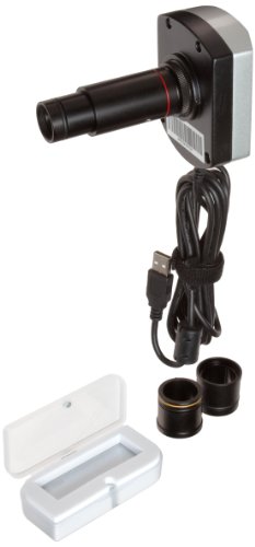
The Ample Scientific TCA-3.0C microscope camera pack has a digital, 3.0 megapixel (MP), complementary metal-oxide semiconductor (CMOS) camera for microscopes with non-compressed image capturing and recording ability, a C/CS-mount interface, and image-processing TSView software. This digital camera for microscopes enhances object or image resolution, and can display an image on a monitor. It is commonly used for education, medical and scientific research, and for industry documentation.
This microscope digital camera features a high-resolution 3.0 MP CMOS color image sensor to manage noise reduction and image quality. It is used with trinocular ports and eyepieces, and connects to a camera with a 1.8-meter USB 2.0 cable. A 1/2" micron sensor shows 2.8 x 2.8 micron pixels, and provides maximum resolution frame rate output of 8 frames per second (FPS) for 2048 x 1536 (horizontal by vertical measurement), and 30 FPS for 640 x 480. The camera supports both automatic and manual adjustment capabilities. It has a TCN 0.5 reduce lens with 23, 30, and 30.6 mm direct insert interface adaptors. The camera has a red, green, and blue (RGB) Bayer filter for color image acquisition. It has an 8-bit RGB pixel mode for depth and image display. Program controls include image size, gain, brightness, exposure time, and color. It has program image processing capabilities of crop, contrast, mirror, invert, rotate, flip, length, area, and angle. In addition to the TCN 0.5 reduce lens, the camera comes with a 0.01 mm micrometer, and a driver installation CD. Included software is compatible with Microsoft Windows 2000, Windows XP, Windows Vista, and Windows 7.
Specifications
| Camera | Digital, 3.0 MP CMOS |
| Interface | C/CS-mount |
| Software | TSView software compatible with Microsoft Windows 2000, Windows XP, Windows Vista, and Windows 7 |
| Camera connection | 1.8-meter USB 2.0 cable |
| Micron sensor | 1/2 inch |
| Pixels | 2.8 x 2.8 micron pixels |
| Maximum resolution frame rate output | 8 FPS for 2048 x 1536 (W x H), 30 FPS for 640 x 480 |
| Data transport and electrical input | 1.8-meter USB 2.0 cable port |
| Lens | TCN 0.5 reduce lens, with 23, 30, and 30.6 mm direct insert interface |
| Filter | RGB Bayer |
| Pixel mode | 8-bit RGB |
| Program control | Image size, gain, brightness, exposure time, and color |
| Program image processing capabilities | Crop, contrast, mirror, invert, rotate, flip, length, area, and angle |
| Micrometer | 0.01mm (included) |
| Operational temperature | 0 to 60 degrees C |
| Storage temperature | -20 to +70 degrees C |
| Operational humidity | 45 to 85% |
| Sensitivity | 1.0V/Lux-S (550nm) |
| S/N ratio | 44dB |
| Dynamic range | >71dB |
| Exposure time | 1ms~0.3s |
| Data interface | 480 megabytes per second (MBps) |
| Mount | Standard C/CS-mount |
| Overall dimensions | 100 x 81 x 48 mm/3.94 x 3.19 x 1.89 inches (H x W x D) |
| Weight | 480g |
H is height, the vertical distance from lowest to highest point; W is width, the horizontal distance from left to right; D is depth, the horizontal distance from front to back.
Microscopes are instruments used to enhance the resolution of an object or image. Types include compound, stereo, or digital. Compound microscopes use a compound optical system with an objective lens and an eyepiece. Stereo microscopes show object depth in a three-dimensional image. Digital microscopes are used to display an image on a monitor, rather than looking through a lens. Microscopes can have monocular (one), binocular (two), or trinocular (three) eyepieces, with varying viewing abilities. A trinocular eyepiece is used to output digital images to an internal or external device such as a camera or monitor. Magnification ability refers to the size of an image. Resolution, also known as resolvant power, refers to the clarity of the image. The interaction between field of view (FOV), numerical aperture (NA), and working distance (WD) determines resolution. Microscopes can control magnification through a fixed focus, or through a range of adjustments. They can also use LED, fluorescent, and mirror light sources to help control viewing capabilities. Microscopes are widely used in education, lab research, biology, metallurgy, engineering, chemistry, manufacturing, and in the medical, forensic science, and veterinary industries.
Ample Scientific manufactures microscopes, centrifuges, and other lab equipment. The company, founded in 1985, is headquartered in Norcross, GA.
What’s in the Box?
- TCA-3.0C CMOS Camera
- TSView software
- TCN-0.5 lens
- 0.01 mm micrometer
- Driver installation CD
- Instructions


No comments:
Post a Comment